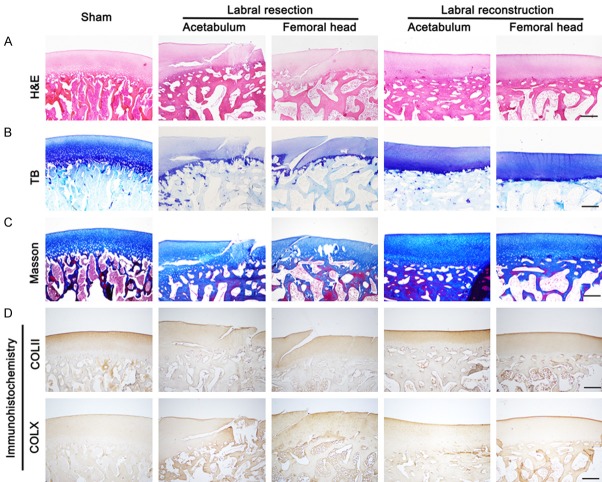
Figure 2.
Histological assessment of articular cartilage of acetabulum and femoral head in sham-operated, resected, and reconstructed joints of porcine models at 24 weeks postoperatively, including H&E, TB, Masson staining, and immunohistochemical staining for type II and X collagen, as shown in (A-D) (sham, n = 3; resection and reconstruction, n = 6). Scale bar, 500 μm.

