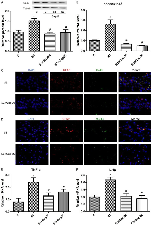
Figure 5.
Gap26 treatment downregulates Cx43 presences and decreases mRNA levels of TNF-α and IL-1β in the hippocampus of surgery mice. A. Representative western blots and densitometric quantification of Cx43 protein. B. Quantitative real-time PCR of Cx43 mRNA expression. C. Protein presence Cx43 in the hippocampus on day 1 treated with (lower panel) or without (upper panel) Gap26. In immunofluorescence images, the nuclei was stained with DAPI-stained (blue), the astrocyte was stained with GFAP (red), and Cx43 was stained in red, respectively. D. Protein presence phosphorylated Cx43 s368 in the hippocampus on day 1 treated with (lower panel) or without (upper panel) Gap26. In immunofluorescence images, the nuclei was stained with DAPI-stained (blue), the astrocyte was stained with GFAP (red), and Cx43 was stained in red, respectively. E. TNF-α mRNA expression in the hippocampus of surgery mice. F. IL-1β mRNA expression in the hippocampus of surgery mice. c: control; s: surgery. N=6. *P < 0.05 versus the controls. #P < 0.05 versus S1.

