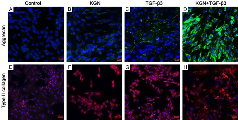
Figure 5.

Immuno-staining of chondrocyte marker proteins in the induced cells. Confocal images of immunofluorescence staining with AGG antibodies (A-D, green) and COL II antibodies (E-H, red) are shown, and the cell nuclei were labeled with DAPI (blue). Original magnification: 20× for (A-D) (scale bar is 20 μm), 10× for (E-H) (scale bar is 50 μm).
