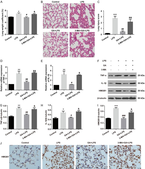
Figure 5.
GA inhibits lung inflammatory injury through autophagy activation in vivo. (A) The pulmonary edema was evaluated by the lung weight coefficient. (B) The histopathological changes of lung tissues were examined using H&E staining (magnification ×400), and (C) morphological damage score for the lung tissues. (D, E) The mRNA levels of TNF-α and IL-1β detected using qRT-PCR. (F) The levels of TNF-α, IL-1β, and HMGB1 were assessed by Western blotting. (G-I) Quantitative analysis of TNF-α, IL-1β, and HMGB1 were shown in bar graphs. (J) The representative pictures of immunohistochemical analysis of HMGB1 protein expression. The experiments were performed four independent times (n=4) and bars represented as mean ± SEM. * P < 0.05, ** P < 0.01, *** P < 0.001 compared with control group. # P < 0.05, ## P < 0.01, ### P < 0.001 in comparison to LPS group. &P < 0.05, &&P < 0.01 versus the GA + LPS group.

