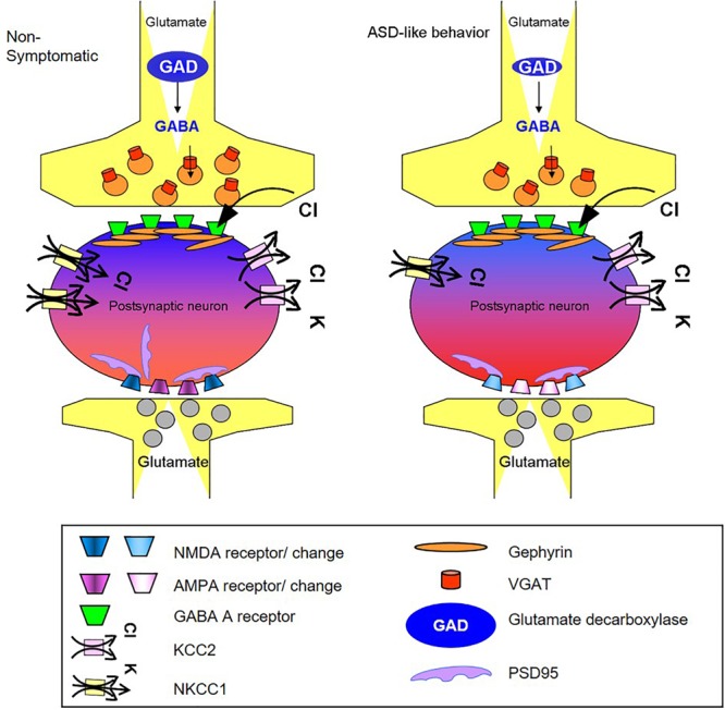
FIGURE 5.

Schematic summary of changes in GABA and glutamate synapse components obtained in the cortex of mice presenting the ASD-like phenotype. The illustration shows the possible molecular origin of the neuronal perturbation that leads to ASD-like behavior in Mthfr deficient mice. The cellular consequences of the changes in GABAergic and glutamatergic proteins observed in the current study: the GABAergic pre-synapse in the ASD-like cortex contains a lower number of vesicular GABA transporters (red) and lower levels of GAD (blue) compared to non-symptomatic cortex. The post-synapse site in the neuron of the ASD-like cortex contains a lower number of NKCC1 transporters (yellow), and thus, GABA receptor (green) activation may result in a less hyperpolarized potential compared to the non- symptomatic cortex (represented by lighter blue color of the neuron in the GABAergic synapse region). The glutamatergic synapse in the cortex of ASD-like mice had a smaller number of PSD-95 molecules (purple). Furthermore, the subunit compositions of the AMPA (purple) and NMDA (blue) receptors in these mice differed from those in the non-symptomatic mice, represented in the figure by the different colors of the receptors. AMPA and NMDA were affected in a sex dependent manner that is not represented in the figure.
