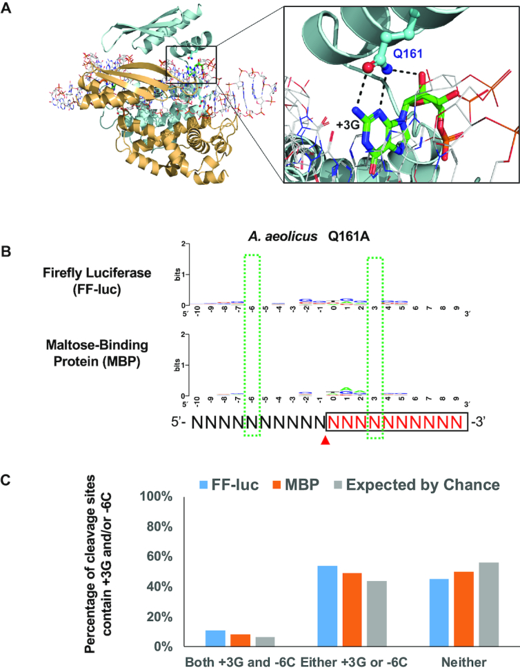
Figure 4.

Residue Q161 of AaRNase III recognizes the +3G near the cleavage site. (A) On the left: schematic illustration of the crystal structure of AaRNase III in complex with dsRNA (PDB entry: 2EZ6); On the right: residue Q161 recognizes the +3G by forming two base-specific hydrogen bonds and one hydrogen bond with the 2′-hydroxyl group. (B) The consensus sequence of cleavage site is abolished when residue 161 was mutated from Q to A. Sequence logos were created as illustrated in Figure 3. (C) Percentage of highly preferable cleavage sites of the AaRNase III Q161A mutant, with or without +3G and/or -6C, is similar to that expected by chance.
