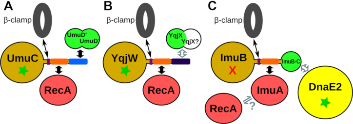
Figure 10.

Schematic comparison of known and proposed complexes and interactions of three mutasomes: (A) Escherichia coli PolV, (B) Bacillus subtilis YqjW (also represents UvrX) and (C) Mycobacterium tuberculosis ImuA-ImuB-DnaE2. Components with similar structures are shown in the same color. Wide black arrows indicate either known interactions or interactions predicted with high confidence and supported by structural models, gray-contoured arrows indicate putative interactions without support of structural models, and narrow black arrows denote transient interactions with the β-clamp. Green star and red X indicate correspondingly the presence and the absence of the polymerase active site.
