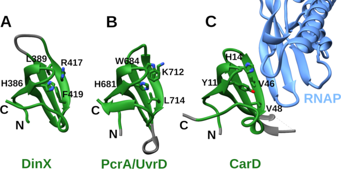
Figure 9.

Comparison of Tudor domains from three proteins: (A) Modeled Tudor-like domain of Mycobacterium tuberculosis DinX; (B) Tudor domain of Geobacillus stearothermophilus PcrA/UvrD helicase (PDB ID: 5DMA); (C) Tudor domain of CarD bound to RNAP β subunit (light blue) (PDB ID: 4KBM). Common parts are highlighted in green. Conserved residues determined to be important for PcrA/UvrD binding to RNAP are labeled and their side chains are shown. Side chains of residues in corresponding positions of other structures are also shown.
