Figure 2.
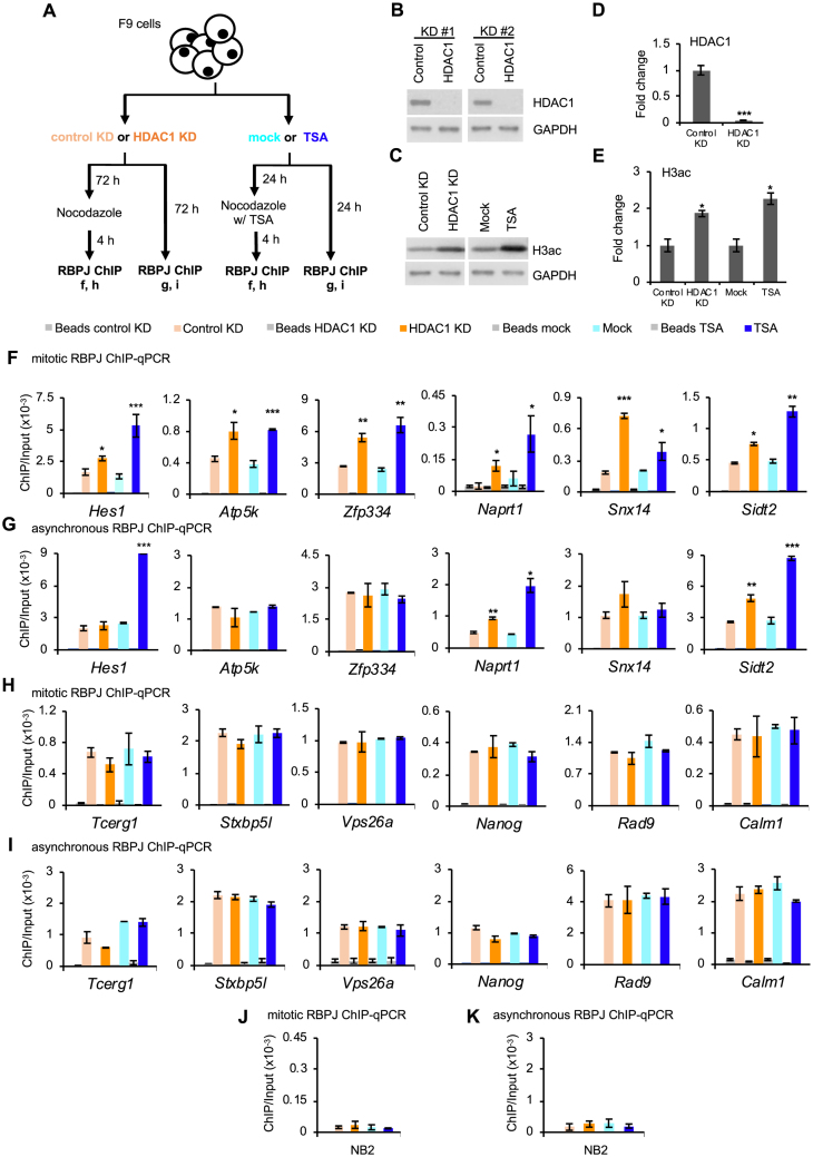
RBPJ occupancy in mitotic cells is negatively regulated by HDAC1 at sites containing the RBPJ-binding consensus. (A) Experimental scheme. Both asynchronous and mitotic cells were collected under the indicated experimental conditions and subjected to RBPJ ChIP-qPCR analysis. (B–E) Representative immunoblots showing relative HDAC1 and histone H3 acetylation (H3ac) abundance (B, D) and immunoblot quantification (C, E). (F–G) RBPJ ChIP analyses at loci that contain the RBPJ-binding motif. (F) RBPJ ChIP-qPCR in mitotic cells. (G) RBPJ ChIP-qPCR in asynchronous cells. (H–I) RBPJ ChIP analyses at loci that do not contain the RBPJ-binding motif (H) RBPJ ChIP-qPCR in mitotic cells. (I) RBPJ ChIP-qPCR in asynchronous cells. (J) Control RBPJ ChIP assay from mitotic cells analysing RBPJ enrichment at an RBPJ nonbinding region (NB3). (K) Control RBPJ ChIP assay from asynchronous cells used to analyse RBPJ enrichment at an RBPJ nonbinding region (NB2). Shown are means ± SEM from two biological replicates. Paired t-tests were performed to compare RBPJ enrichment in treated relative to untreated cells. *P ≤ 0.05; **P ≤ 0.01; ***P ≤ 0.001.

