Figure 6.
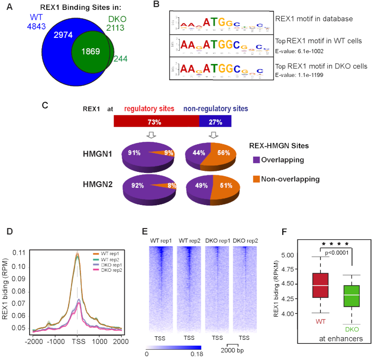
Loss of HMGNs leads to a global reduction in REX1 binding to chromatin. (A) Venn diagram showing the number of REX1 binding sites detected by ChIP-seq in WT and DKO ESCs. (B) Top DNA sequence motif underlying the REX1 binding sites in WT and DKO cells. (C) Overlap between REX1 and HMGNs occupancy at chromatin regulatory sites. (D, E) Decreased REX1 occupancy at TSS and neighboring 4 kb regions in DKO cells. (F) Box plots showing decreased REX1 binding at enhancers of DKO cells. ****P < 0.0001.

