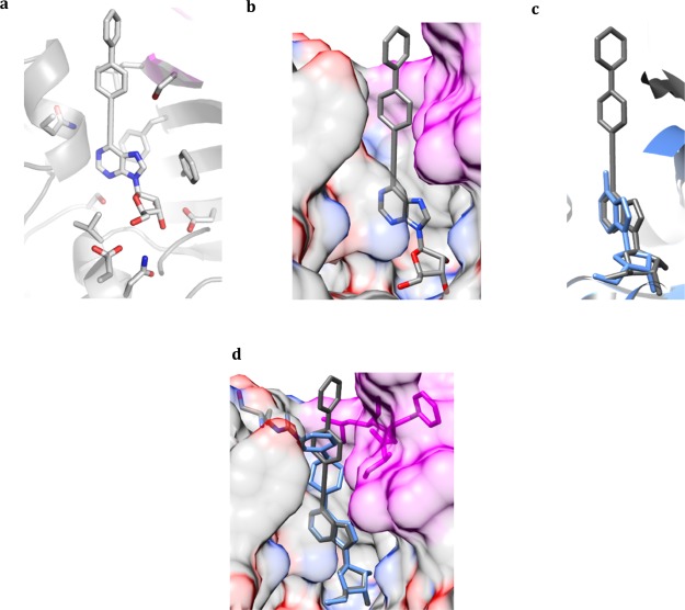
Figure 6.
Crystal structure of the MtbAdoK-17 complex. (a) 17 bound to the active site of MtbAdoK. (b) The large bulky substitution gets accommodated in the “chimney-like” cavity. (c) Compound 17 (gray) binds in a different orientation with respect to adenosine (blue). (d) Superimposition of crystal structure complexes of 17 (gray) and 7 (blue). Chain B residues forming the distal part of the “chimney-like” cavity colored magenta while chain A residues are colored by heteroatom.

