
Figure 5.
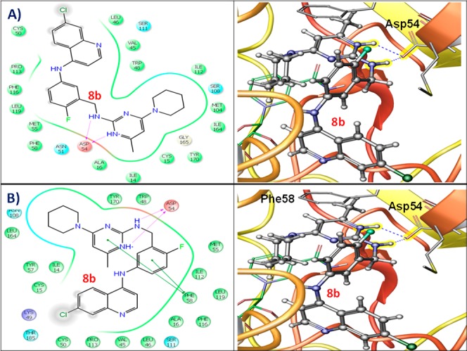
Two- and three-dimensional docking poses of compound 8b showing the interactions in the binding sites of (A) wild type Pf-DHFR-TS (PDB ID: 3QGT) and (B) quadruple mutant Pf-DHFR-TS (PDB ID: 3QG2).
Official websites use .gov
A
.gov website belongs to an official
government organization in the United States.
Secure .gov websites use HTTPS
A lock (
) or https:// means you've safely
connected to the .gov website. Share sensitive
information only on official, secure websites.
Two- and three-dimensional docking poses of compound 8b showing the interactions in the binding sites of (A) wild type Pf-DHFR-TS (PDB ID: 3QGT) and (B) quadruple mutant Pf-DHFR-TS (PDB ID: 3QG2).