Figure 2.

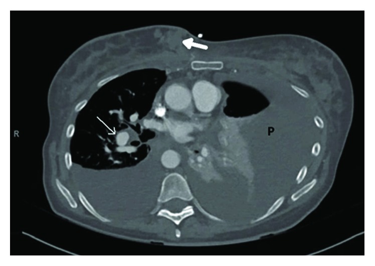
Computed tomography with angiography showing a right medial breast mass (closed white arrow) with mediastinal (open white arrow) and axillary lymphadenopathy. Large effusions greater on the left (P).

Computed tomography with angiography showing a right medial breast mass (closed white arrow) with mediastinal (open white arrow) and axillary lymphadenopathy. Large effusions greater on the left (P).