Figure 3.

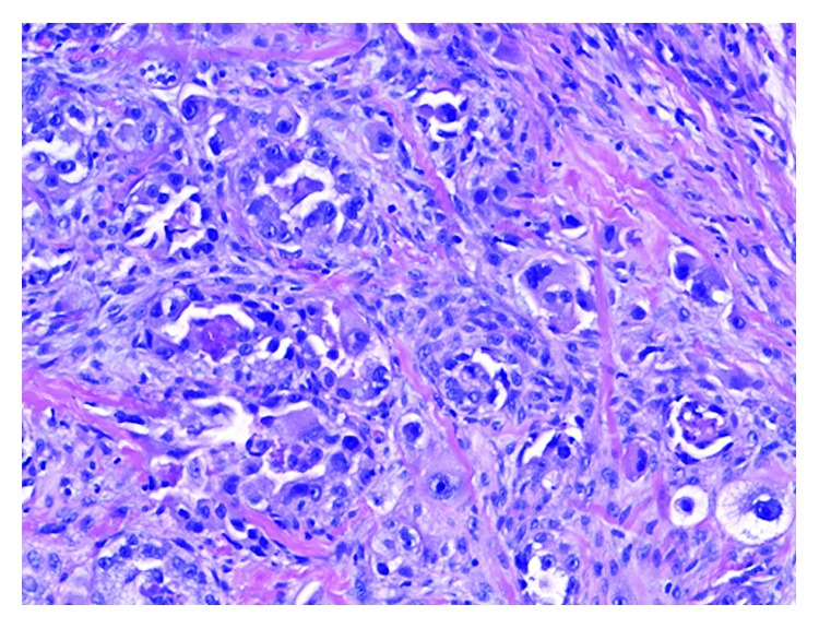
Hematoxylin and eosin stain showing invasive carcinoma consisting of pleomorphic tumor cells with vacuolated and eosinophilic cytoplasm and prominent nucleoli.

Hematoxylin and eosin stain showing invasive carcinoma consisting of pleomorphic tumor cells with vacuolated and eosinophilic cytoplasm and prominent nucleoli.