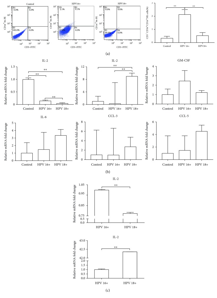
Figure 1.
The differences of NK cells between HPV16- and HPV18-infected cervixes. (a) Distribution of CD56+CD16+ NK cells was analyzed in the cervical brush specimens by FACS. (b) Several soluble cytokines that represent NK cell involvement in the immune status were investigated in the cervical brush specimens by quantitative real-time PCR. (c) IFN-γ and IL-2 expressions were measured in the cervical conization tissue by quantitative real-time PCR. ∗P < 0.05; ∗∗P < 0.01.

