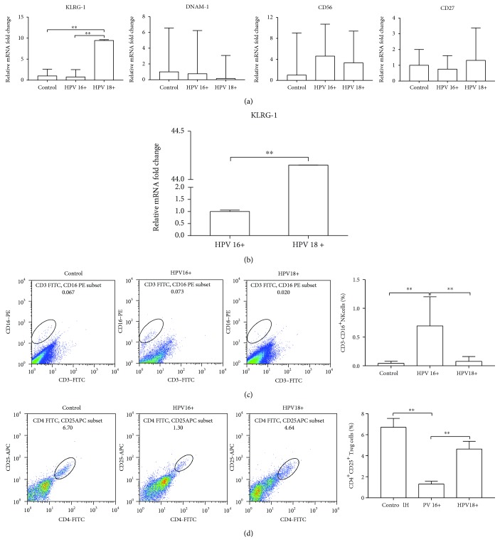
Figure 2.
The different effects of NK cells between HPV16- and HPV18-infected cervixes. (a) Four typical cell membrane markers of NK cells were evaluated in the cervical brush specimens by quantitative real-time PCR. (b) KLRG-1 expression was measured in the cervical conization tissue by quantitative real-time PCR. (c) Distribution of CD16+ NK cells was analyzed in the cervical brush specimens by FACS. (d) Distribution of CD4+CD25+ Treg cells was analyzed in the cervical brush specimens by FACS. ∗P < 0.05; ∗∗P < 0.01.

