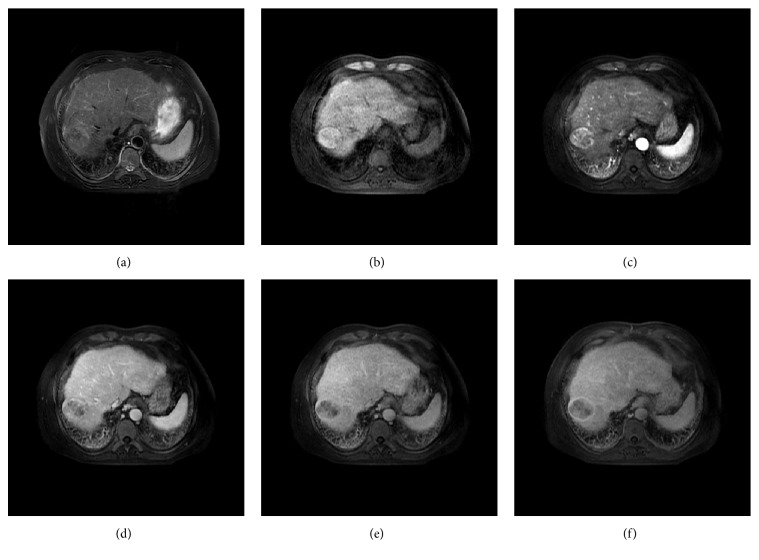
Figure 4.
Axial MR images and pathologic image of a 52-year-old man with HCC. (a) A fat-suppressed T2-weighted fast spin-echo image shows an oval-shaped, heterogeneous, slightly hyperintense neoplasm in segment VII with a maximum diameter of 4.6 cm. Axial precontrast (b), late artery phase (c), portal vein phase (d), equilibrium phase (e), and delay phase (f) T1-weighted 3D GRE images show a hyperintense appearance of the lesion on precontrast T1-weighted images (b), obvious enhancement in the late arterial phase (c), and washout in the portal vein phase (d); an enhancing capsule was detectable in the equilibrium phase (e) and was more obvious in the delay phase (f); all of these MRI features are consistent with typical HCC. The tumor was successfully surgically resected and was pathologically confirmed as an MD HCC with no vascular invasion.

