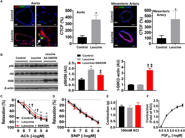
Figure 1.

Leucine‐induced activation of mTORC1 impairs endothelial‐mediated relaxation. A, Representative images of aortic and mesenteric arterial rings cultured for 24 hours in leucine‐supplemented (10 mmol/L) media compared with control (0.45 mmol/L) media. Phospho‐S6 (pS6; red) denotes mTORC1 signaling and Von Willebrand Factor (VWF, green) staining denotes the endothelium. White arrow denotes co‐localization of pS6 and Von Willebrand Factor staining. Images taken from 3 to 4 independent experiments and quantification data of aortic and mesenteric arterial rings expressed as percentage corrected total cell fluorescence (CTCF%) are shown. B, Representative Western blot images of aortic rings cultured in control, leucine‐supplemented and leucine‐supplemented+Ad‐S6KDN (2×10⁸ pfu/mL) media for phospho‐S6, total S6, total S6 kinase and β‐actin. Quantification data are expressed as arbitrary units (n=6–9/group). Vascular reactivity responses of cultured aortic rings to (C) endothelial‐dependent acetylcholine, (D) endothelial‐independent sodium nitroprusside (SNP), and contractile responses to (E) 100 mmol/L potassium chloride (KCl) and (F) prostaglandin F2α (PGF2α) (n=10/group). ACh indicates acetylcholine; CTCF %, percentage corrected total cell fluorescence; mTORC1, mechanistic target of rapamycin complex 1; SNP, sodium nitroprusside; *P<0.05 leucine vs control; † P<0.05 leucine vs leucine+S6KDN, ‡P<0.05 control vs leucine+S6KDN.
