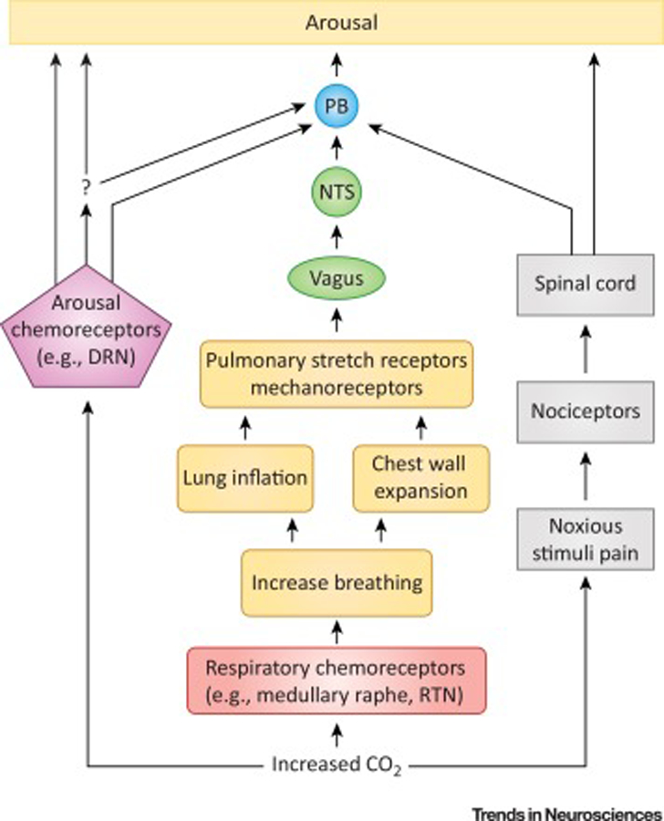
Figure 2. Schematic depicting possible mechanisms for CO2 induced arousal.

Inspired CO2 rapidly induces a rise in serum CO2 concentration (bottom), which is detected by peripheral (e.g. carotid bodies; not depicted) and central respiratory (red) and arousal (purple) chemosensors. Evidence implicates medullary raphe 5-HT neurons and glutamatergic RTN neurons as important respiratory chemosensors. In a long-standing proposed mechanism for CO2 arousal (middle) activation of respiratory chemosensors increases breathing, causing lung inflation and chest wall expansion, which activates stretch and mechanoreceptors in these structures (orange). This activation is transmitted via the vagus nerve to the NTS (green) and ultimately to PB (blue) which leads to arousal (yellow). The PB may be the final common mediator of arousal to a number of different stimuli. Arousal pathways downstream of PB were analyzed by Kaur et al. [45] and not depicted here. In the more recently emerging proposed CO2 arousal mechanism (left), changes in serum CO2 are detected directly by arousal chemosensors which cause arousal, perhaps through a PB mediated mechanism, either directly, or indirectly through some yet to be determined intermediary (question mark). A leading candidate for the arousal chemosensors is DRN 5-HT neurons; however, other structures such as norepinephrine (NE) neurons in LC and orexin neurons in the lateral hypothalamus (LH) have been implicated. In addition, the NTS is itself chemosensitive. There can also be a noxious component to inspiration of CO2 which can cause arousal through spinally mediated nociceptive pathways (right; black). DRN, dorsal raphe nucleus; NTS, nucleus tractus solitarius; PB, parabrachial nucleus; RTN, retrotrapezoid nucleus
