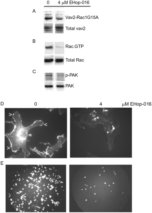
Figure 6.5.
Effect of EHop-016 on Rac activation and downstream activities in highly metastatic MDA-MB-435 cells. (A) GST-Rac1(G15A) (mutant form of Rac that selectively binds active GEFs) beads were preincubated with vehicle (0), or 4 μM EHop-016 prior to incubation with MDA-MB-435 cell lysates. A representative western blot (N = 3) immunostained for Vav2 is shown. Top row, pulldown; bottom row, total cell lysate. (B) MDA-MB-435 cells treated with vehicle (0) or EHop-016 (4 μM) for 24 h were lysed and subjected to a pulldown assay for Rac.GTP using a GST-CRIB domain of PAK and western blotted with a pan Rac antibody (Rac1,2,3). Representative western blot (N = 3) is shown. Top row, pulldown; bottom row, total cell lysate. (C) MDA-MB-435 cells were treated with vehicle (0) or 4 μM EHop-016 for 24 h and the cells lysed and western blotted for active P-thr 423 PAK (upper band) or total PAK (lower band). Representative western blot (N = 3) is shown. (D) MDA-MB-435 metastatic breast cancer cells were treated with vehicle or EHop-016 at 4 μM for 24 h. Cells were fixed, permeabilized, and stained with Rhodamine phalloidin to visualize F-actin. Representative micrographs are shown. Arrows indicate lamellipodia. (E) MDA-MB-435 cells treated with vehicle (0) or EHop-016 (4 μM) for 24 h were subjected to a Transwell migration assay. The cells that migrated to the underside of the membrane of the top well (through 8 μM diameter pores) were stained with propidium iodide and quantified. Representative micrographs of cells that migrated for each treatment are shown.

