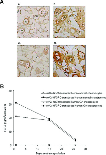
Figure 1.

FGF‐2 expression in vitro. (A) Immunocytochemical detection of FGF‐2 in spheres carrying human normal (a and b) and OA chondrocytes (c and d) after 26 days. Cells were transduced with rAAV‐lacZ (a and c) or rAAV‐hFGF‐2 (b and d), encapsulated, and processed after 26 days (n= 9 per condition) to detect FGF‐2 (1:50). Magnification ×100. (B) Time course analysis of FGF‐2 production in culture supernatants from spheres. Conditioned medium was collected at the denoted time points after encapsulation (n= 9 per time point and condition) and FGF‐2 secretion was measured by ELISA (±S.D.).
