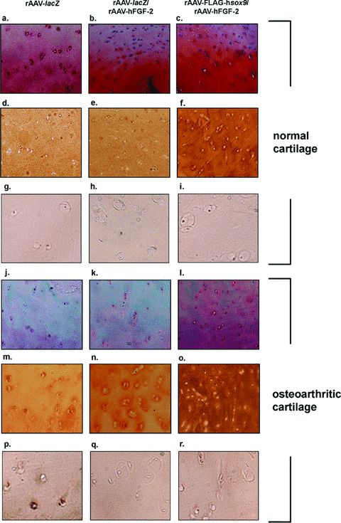
Figure 5.

Safranin O staining and type‐II and type‐X collagen immunostaining in co‐transduced cartilage. Human normal (a–i) and OA cartilage (j–r) were transduced with rAAV‐lacZ (a, d, g, j, m and p), rAAV‐lacZ/rAAV‐hFGF‐2 (b, e, h, k, n, and q), or rAAV‐FLAG‐hsox9/rAAV‐hFGF‐2 (c, f, i, l, o and r) and processed after 10 days (n= 6 per condition) for staining by safranin O and haematoxylin and eosin (a–c and j–l, magnification ×40) and to detect type‐II collagen (1:50) (d–f and m–o, magnification ×40) and type‐X collagen (1:200) (g–i and p–r, magnification ×200). Representative view of the middle zone.
