Figure 7.

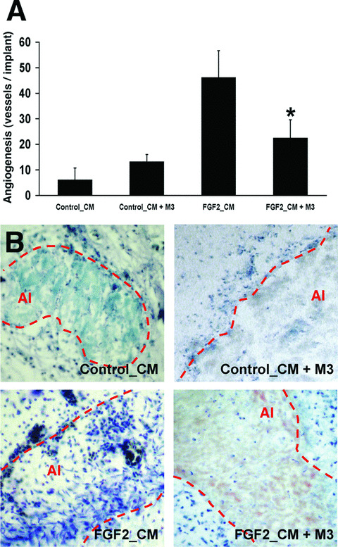
The conditioned medium (CM) from FGF2‐stimulated microvascular endothelial cells promotes chemokine‐dependent angiogenesis in vivo. (A) Chick embryo CAM assay was performed with the CM from non‐stimulated (Control_CM) and FGF2‐stimulated (FGF2_CM) 1G11 endothelial cells in the absence or presence of 75 ng of the pan‐chemokine inhibitor M3. Data are expressed as the mean ± SD of the number of vessels invading the alginate area (*, statistically different from the ‘FGF2_CM’ group, P < 0.05). (B) Representative histological sections of CAMs from the different experimental groups (May Grünwald‐Giemsa staining). Note that FGF2_CM induces neovascularization and a strong inflammatory cell infiltrate within the alginate implant (AI, dotted line), both greatly reduced in the presence of M3.
