
Figure 1.
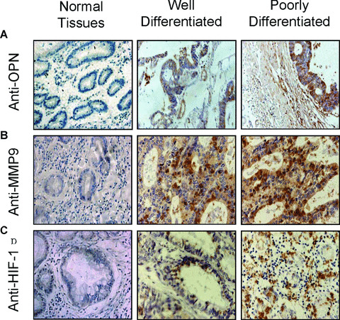
Immunohistochemical staining of OPN, MMP9 and HIF‐1α in human gastric cancer tissues. The tissue sections of the primary gastric cancers, including the well‐ and poorly differentiated gastric cancer tissues, and the matched non‐cancer gastric tissues, were immunostained with anti‐OPN (A), anti‐MMP9 (B) and anti‐HIF‐1α (C) antibodies respectively. The positive staining for OPN, MMP9 or HIF‐1α proteins were shown in brown colour. All sections were counterstained with haematoxylin showing in blue colour (×100).