
Figure 4.
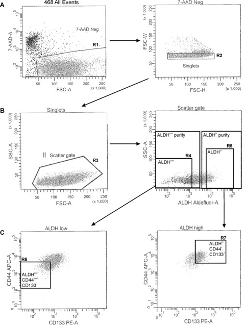
Strategy for isolation of stem‐like human breast cancer cells from the MDA‐MB‐468 cell line. Fluorescence‐activated cell sorting (FACS) was used to isolate ALDHhiCD44+ and ALDHlowCD44low/− human breast cancer cells for functional assays. MDA‐MB‐468 cells were concurrently labelled with 7‐AAD, fluorescent antibodies (CD44‐APC + CD133‐PE) and the ALDEFLUOR™ assay kit. Cell subsets were isolated using a four‐colour protocol on a FACS Vantage/Diva cell sorter, including ALDHhiCD44+CD133+ and ALDHlowCD44low/−CD133− subsets (A–C): Representative schematic of a sequentially gated MDA‐MB‐468 cell line sort. (A) Cells were first selected for viability based on 7‐AAD negativity (R1, left panel) and for singlets (R2, right panel). (B) Cells were then selected based on light scatter (R3, left panel) and divided into ALDHlow (R4, bottom ∼20% of parent population) and ALDHhi (R5, top ∼20% of parent population) based on ALDH activity (right panel). (C) Finally, ALDHlow cells were further selected based on a CD44low/−CD133low/− phenotype (R6, bottom 10% of parent population, gated on R1 + R2 + R3 + R4) (left panel), whereas ALDHhi cells were further selected based on expression of a CD44+CD133+ phenotype (R7, top ∼10% of parent population, gated on R1 + R2 + R3 + R5) (right panel). The resulting cell subsets were designated as ALDHhiCD44+CD133+ (R7, ‘stem‐like’) and ALDHlowCD44low/−CD133− (R6, non ‘stem‐like’), and were collected for functional analysis of differences in malignant and metastatic behaviour in vitro and in vivo.