
Figure 5.
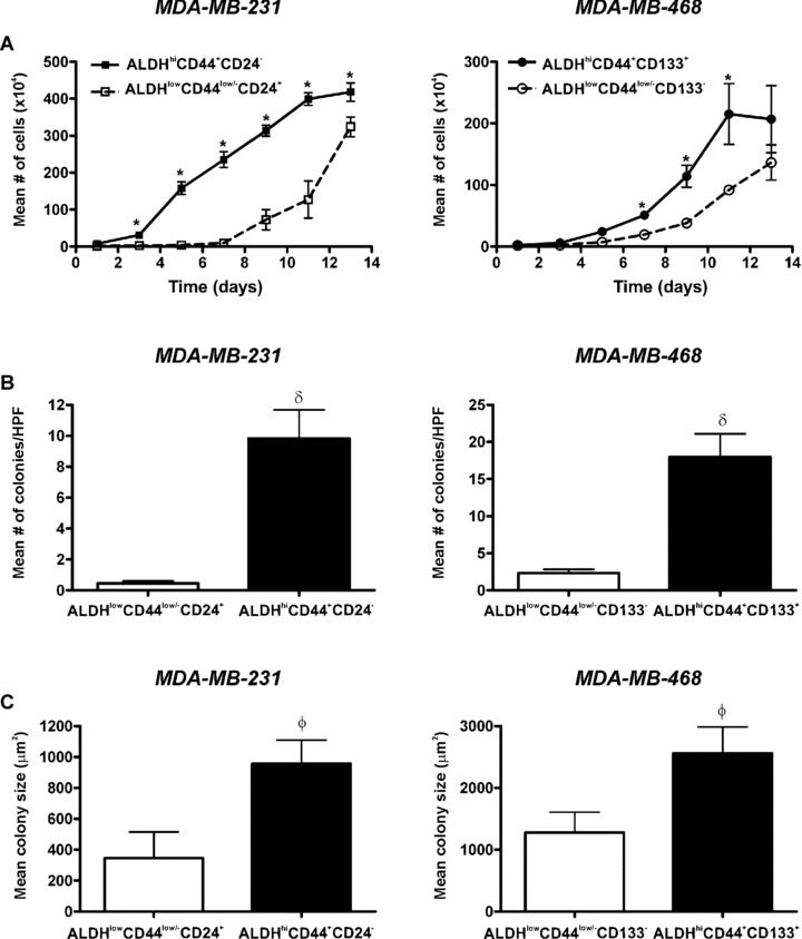
ALDHhiCD44+CD24− and ALDHhiCD44+CD133+ breast cancer cells demonstrate enhanced cell growth and colony formation in vitro. Cells were isolated by FACS as described in Figs 3 and 4 and subjected to in vitro assays for growth and colony formation. (A) Cell growth kinetics in normal (anchorage‐dependent) culture over time of ALDHhiCD44+CD24− (▪) versus ALDHlowCD44low/−CD24+ (□) cells isolated from the MDA‐MB‐231 cell line (left panel); and ALDHhiCD44+CD133+ (•) versus ALDHlowCD44low/− CD133− (○) cells isolated from the MDA‐MB‐468 cell line (right panel) (5.0 × 10⁴ cells/60 mm plate; n = 3 plates/time‐point). Data are presented as the mean ± S.E.M. *= significantly different cell number than respective ALDHlowCD44low/− cell subsets (P < 0.05). (B, C) Colony forming ability of ALDHhiCD44+CD24− (MDA‐MB‐231) and ALDHhiCD44+CD133+ (MDA‐MB‐468) cells (black bars) versus respective ALDHlowCD44low/− cell subsets (white bars) isolated from MDA‐MB‐231 (left panels) or MDA‐MB‐468 (right panels) cell lines. Cells (1.0 × 10⁴ cells/60 mm plate, n= 4 plates/cell population) were grown in soft agar (0.6%) for 4 weeks. Five high‐powered fields of view (100×) were counted for each dish. (B) Mean number of colonies per plate and (C) Mean colony diameter (μm2). Data are presented as the mean ± S.E.M. δ= significantly different colony number than respective ALDHlowCD44low/− cell subsets (P < 0.001). The value φ= significantly different colony size than respective ALDHlowCD44low/− cell subsets (P < 0.05).