
Figure 7.
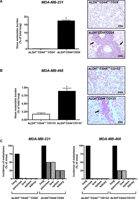
ALDHhiCD44+CD24− and ALDHhiCD44+CD133+ breast cancer cells demonstrate enhanced metastatic growth in vivo following tail vein injection. Cells were isolated by FACS as described in Figs 3 and 4 and injected into the lateral tail vein of female NOD/SCID‐IL2Rγ null mice using an established model of experimental metastasis (5 × 10⁵ cells/mouse; 4 mice/cell population). At 5 weeks (MDA‐MB‐231) or 12 weeks (MDA‐MB‐468) after injection, mice were killed and assessed for metastatic burden in the lung and elsewhere. Tissue sections were subjected to haematoxylin and eosin staining (five random sections/tissue/mouse), and the incidence and extent of metastasis was determined in a blinded fashion. (A and B) Quantitative analysis of tumour burden (mean% of lung occupied by tumour) (left panels) and representative haematoxylin and eosin stained lung sections (right panels) following tail vein injection of (A) ALDHhiCD44+CD24− versus ALDHlowCD44low/−CD24+ cells isolated from the MDA‐MB‐231 cell line; and (B) ALDHhiCD44+CD133+ versus ALDHlowCD44low/−CD133− cells isolated from the MDA‐MB‐468 cell line. Arrowheads on haematoxylin and eosin images indicate regions of tumour within the lung. Data are presented as the mean ± S.E.M. *= significantly different than respective ALDHlowCD44low/− subsets (P < 0.05). (C) Incidence of metastatic growth in lung and extrapulmonary tissues following tail vein injection of ALDHhiCD44+CD24− versus ALDHlowCD44low/−CD24+ cells isolated from the MDA‐MB‐231 cell line (left panels); and ALDHhiCD44+CD133+ versus ALDHlowCD44low/−CD133− cells isolated from the MDA‐MB‐468 cell line (right panels).