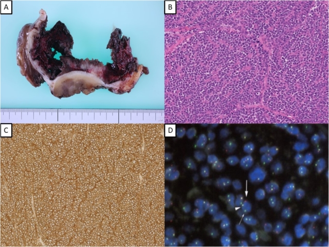
Fig. 2.

(A) The macroscopic specimen showed hematoma in the thickened cyst wall. (B) Microscopic examination of the hematoxylin-eosin-stained sample showed uniform small round blue cells arranged as a rosette fashion ( × 400). (C) Immunohistochemistry was positive for CD99 ( × 400). (D) Fluorescence in situ hybridization analysis was positive for the EWSR1 gene in 90% of cells.
