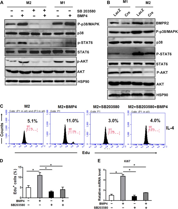
Figure 4.
Activation of M2 macrophage is regulated by the p38/MAPK/STAT6/PI3K–AKT pathway. (A) Representative western blot images of the expression levels of molecules involved in the BMP signalling pathway in M2 and M1 macrophages treated with or without BMP4 (25 ng/ml) for 1 h. For p38 inhibition, cells were pretreated with p38 inhibitor SB203580 (20 nM) for 6 h. (B) Representative western blot images of molecules involved in the BMP signalling pathway in BMDMs derived from BMPR2LoxP/LoxP genotype mice and knocked down for BMPR2 by expressing Cre recombinase. (C) M2 macrophages were pre-incubated with the p38 inhibitor SB203580 (20 mM) for 6 h before addition of BMP4 (25 mg/ml). The amount of EdU+ macrophages was counted. (D) The quantification of EdU+ cells described in C from three individual experiments. (E) The mRNA levels of Ki67 in different groups of cells described in C. Data were collected from three individual experiments and presented as mean ± SD. Student’s test was used for comparisons. *P < 0.05; **P < 0.01; ***P < 0.001.

