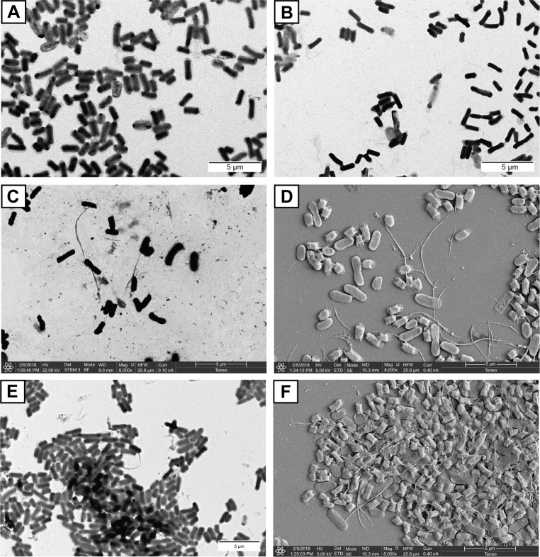
Figure 9.
Representative, negatively stained TEM and sputter coated with gold, micrographs of the interaction of glyconanotubes I and II with E. coli strains ORN178 and ORN208.
Notes: (A) TEM micrograph of E. coli strain ORN178 alone. (B) TEM micrograph of E. coli strain ORN208 alone. (C) TEM micrograph of the lactose-coated glyconanotube II incubated with the E. coli strain ORN178. (D) SEM micrograph of sputter coated with gold of glyconanotube II incubated with E. coli strain ORN178. (E) TEM micrograph of the mannose-coated glyconanotube I incubated with the E. coli strain ORN178. (F) SEM micrograph of sputter coated with gold of glyconanotube I incubated with E. coli strain ORN178. For conditions, see “Materials and methods” section in the text. Size bars are shown.
Abbreviations: TEM, transmission electronic microscopy; SWCNTs, single-walled carbon nanotubes; E. coli, Escherichia coli; SEM, scanning electronic microscopy.

