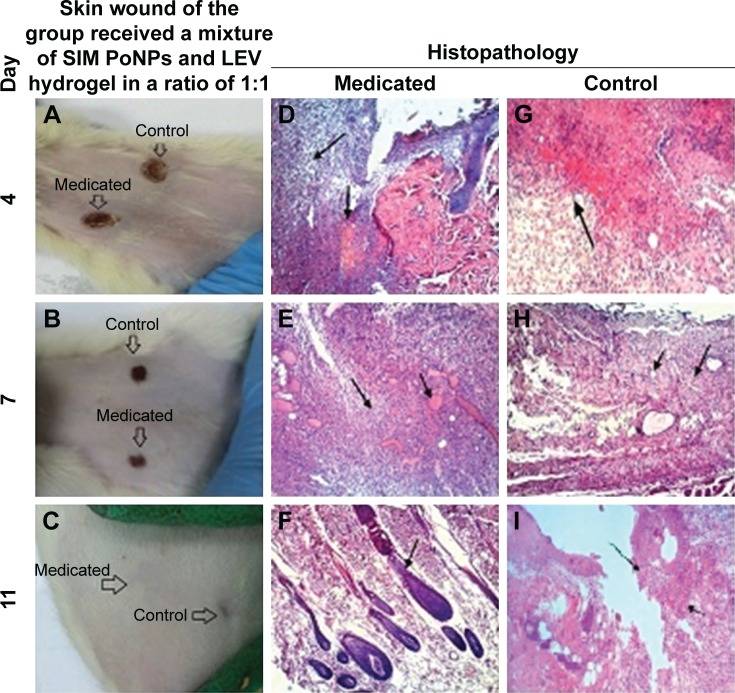
Figure 11.
Skin wound and histopathology of the group received a mixture of SIM PoNPs and LEV hydrogel at a ratio of 1:1.
Notes: (A) No redness or edema appeared around the wound. (B) Medicated wound started to epithelize. (C) Wounds had been completely cured. (D) Focal necrosis (small arrow) and inflammatory cell infiltration (large arrow). (E) Well-vascularized (small arrow) formation of granulation tissue (large arrow). (F) Normal hair follicle formation. (G) Necrosis (small arrow). (H) Well-vascularized (small arrow) granulation tissue formation (large arrow). (I) Incomplete epithelization (arrow) and severe inflam matory cell infiltration in the dermal layer and focal areas of hemorrhage in the subepithelial region (small arrow).
Abbreviations: SIM, simvastatin; LEV, levofloxacin hemihydrate; PoNPs, polymeric nanoparticles.

