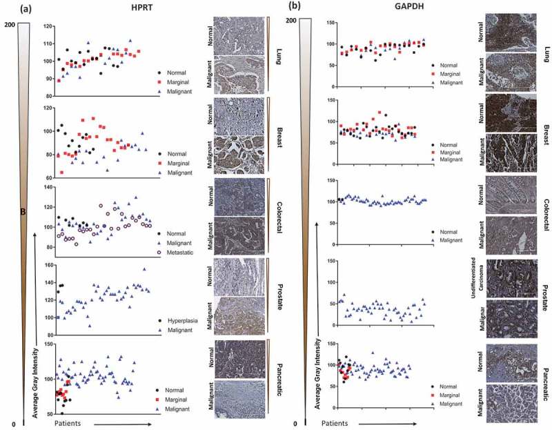
Figure 1.

Immunohistochemistry staining of HPRT compared to GAPDH in a variety of organ types. Lung, Breast, Colon, Prostate, and Pancreatic malignant and normal tissue were stained with antibodies against HPRT and GAPDH to determine trends in expression between cancerous and healthy tissue. Tissues were quantified on a gray scale and lower values indicate a darker stain and higher protein binding. The gradient scale directly left of the images shows the directionality and intensity of the DAB staining. (A) HPRT showed a significant variability between malignant and normal tissue samples with an overall trend of increased HPRT upon malignancy. (B) GAPDH had significantly elevated levels of expression in both malignant and normal tissue samples.
