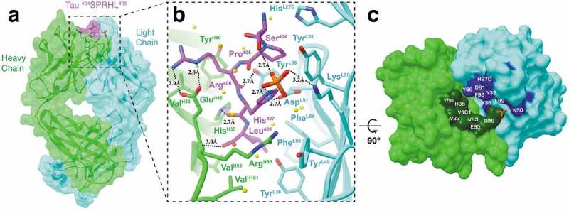
Figure 3.

Fab structure of 8B2 in complex with a tau peptide. (a) Surface representation of the Fab 8B2/peptide complex with underlying ribbon display. Heavy and light chains are colored light green and cyan, respectively. The tau peptide is shown in magenta with the epitope sequence. Although the peptide used in crystallization is 23 residues in length, only five residues (404SPRHL408) are visible in the electron density map. (b) A front view ribbon representation of Fab 8B2 in complex with the non-phosphorylated tau peptide. Key residues involved in antigen-binding and a phosphate observed in the binding site next to the side chain of Ser404 are shown as sticks. Hydrogen bonds are represented by dashed black lines and labeled with bond distances. Water molecules are represented by yellow spheres. (c) Top-down view of the paratope. Surface areas of the contact residues from the heavy chain are shown in dark green and that from the light chain in blue. The phosphate observed at the binding site is shown as a reference. See also Figure S3.
