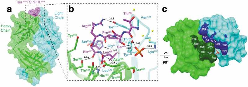
Figure 4.

Fab structure of 6B2 in complex with a tau peptide. (a) Surface representation of the Fab 6B2/peptide complex with underlying ribbon display. Six residues (403TSPRHL408) are visible in the electron density map. (b) A detailed front view of Fab 6B2 in complex with a non-phosphorylated tau peptide. Again, a phosphate is observed next to the side chain of Ser404 at the antigen binding site. (c) Top-down view of the paratope surface.
