
Figure 2.
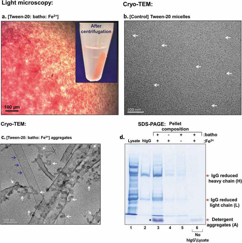
(a) Light microscopy images of Tween-20 aggregates comprising Tween-20 micelles, the hydrophobic chelator bathophenanthroline (batho) and Fe2+ ions. The red color derives from the [(batho)3:Fe2+] complex. The white arrow points to the red pellet generated by brief (14K, 2 min.) centrifugation (inset). (b) and (c). Cryo-TEM images of Tween-20 micelles White arrows point to individual micelles (dark black dots). Tween-20 aggregates as in (a). White arrows point to Tween-20 aggregates, while blue arrows points to an elongated crystal of excess hydrophobic chelator (batho). (d) SDS-PAGE analysis of Tween-20 pellets and process dependence on the chelator (batho) and Fe2+ Lane 1: E. coli lysate – serving as an artificial contamination background; Lane 2: target human (hIgG); Lane 3: pellet composition obtained after incubating Tween-20 aggregates comprising [Tween-20:batho:Fe2+] with a mixture of [E. coli lysate:hIgG], followed by centrifugation and supernatant removal as described in Experimental Section; Lanes 4–5: as in lane 3, but in the absence of batho or of Fe2+, respectively; Lane 6: Detergent aggregates devoid of added protein. H, L denote the reduced heavy and light chains of the target antibody, respectively. A points to the detergent aggregate band, migrating at the front of the gel.