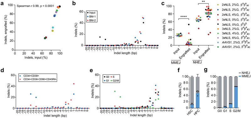
Figure 4 |. Persistence of NHEJ repaired alleles in HSCs.
a), Correlation of indel frequencies of input HSPCs to indel frequencies of engrafted human cells in mice BM after 16 weeks. Each dot represents average indel frequencies of mice transplanted with the same input HSPCs. Legend denoting transplant is same as in (c). The Pearson correlation coefficient (r) is shown. b, Indel spectrum of input cells from healthy donor βAβA#2 electroporated with 2xNLS-Cas9 (coupled with sgRNA-1617) supplemented with 2% glycerol and engrafted 16 week BM human cells. c, Relative loss of edited alleles repaired by MMEJ and gain of edited alleles repaired by NHEJ in mice BM 16 weeks after transplant. The indel spectrum was determined by deep sequencing analysis. Indel length from −8 to +6 bp was calculated as NHEJ, and from −9 to −20 bp as MMEJ. These data comprise 28 mice transplanted with 8 BCL11A enhancer edited inputs and 5 mice transplanted with 2 AAVS1 edited inputs. Median of each group is shown as line, **P < 0.005, ****P < 0.0001 as determined by Kolmogorov–Smirnov test. d-e, Indel spectra of HSPCs stained and sorted 2h after RNP electroporation with 3xNLS-Cas9 with sgRNA-1617. HSPCs prestimulated for 24h prior to electroporation. HSPCs stained with CD34, CD38, CD90, CD45RA in (d) and with Pyronin Y, Hoechst 33342 in (e). Indels determined by Sanger sequencing with TIDE analysis after culturing cells for 4 days after sort. Relative loss of edited alleles repaired by MMEJ and gain of edited alleles repaired by NHEJ at BCL11A enhancer and AAVS1 in sorted enriched HSCs (f) or G0 phase cells (g) shown. Data are plotted as mean ± SD for (f, g) and analyzed using unpaired two-tailed Student’s t tests. Data are representative of three biologically independent replicates.

