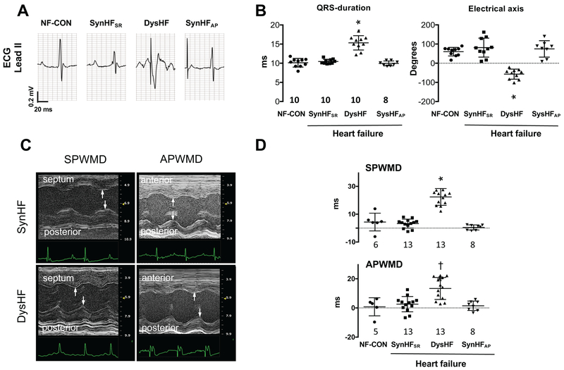
Figure 3. Electromechanical dyssynchrony in mice.
(A) Representative electrical tracings from non-failing mice in sinus rhythm (NF-CON) and after I/R with normal sinus rhythm (SyncHFSR), RV over-drive pacing (DysHF), or atrial over-drive pacing (SyncHFAP). (B) Average QRS-duration and electrical axis for the four groups. (C) M-mode echocardiograms showing increased septum-to-posterior wall motion delay (SPWMD) and anterior-to-posterior wall motion delay (APWMD) in post I/R mice at SR (SyncHFSR) or with RVP (DysHF). The delay increases with RVP. (D) Summary data for both delay indexes. Summary analysis by 1-Way ANOVA, Tukey post-hoc pairwise comparisons: *<0.0001 versus all other groups, † p=0.002 vs NF-CON, p=0.0003 vs SynHFSR, p=0005 vs SynHFAP.

