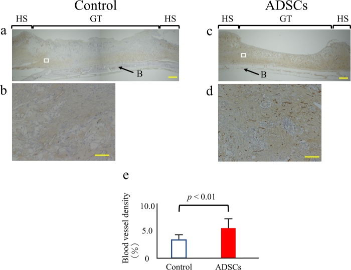
Fig 5. Quantitative analysis of wound neovascularization.
(a-d) Control and ADSC specimens at 14 days after surgery were stained for CD31, a blood vessel endothelial cell marker (brown) (a, c: 40x magnification. Scale bars = 1 mm). For each group, the figure below is a higher-magnification micrograph of the boxed region in the figure above (b, d: 400x magnification. Scale bars = 100 μm). (e) Blood vessel density was calculated by dividing the area of CD31-positive vessels by the total area. The data represent the mean ± SD. HS = healthy skin; GT = granulation tissue; B = cranial bone.

