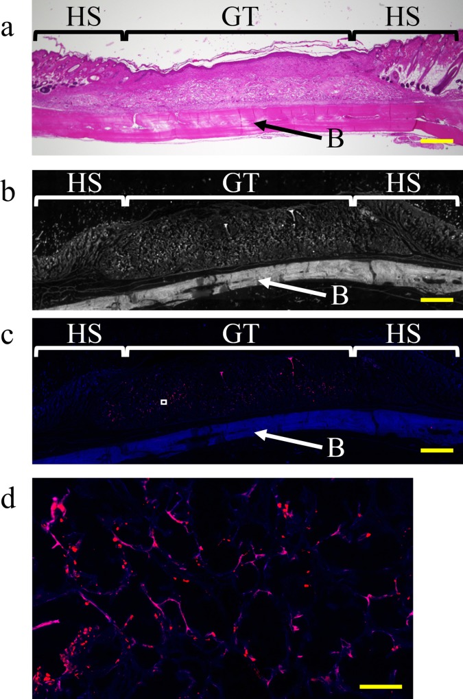
Fig 6. Representative images of DiI labeling of the center of the wound along with implantation time.
Frozen sections were prepared at 21 days after transplantation of ADSCs labeled with DiI dye. (a) Magnified microphotographs of sagittal sections of hematoxylin and eosin-stained specimens collected from the ADSC group 21 days after surgery. (b) Grayscale values of frozen sections of each tissue (40× magnification); these values were similar to those for the frozen section in (c). Scale bar, 1 mm. (c) Frozen section with DiI labeling (40× magnification. Scale bar, 1 mm). (d) High-magnification image (500×) of the boxed region in (c). Scale bar, 100 μm. HS = healthy skin; GT = granulation tissue; B = cranial bone.

