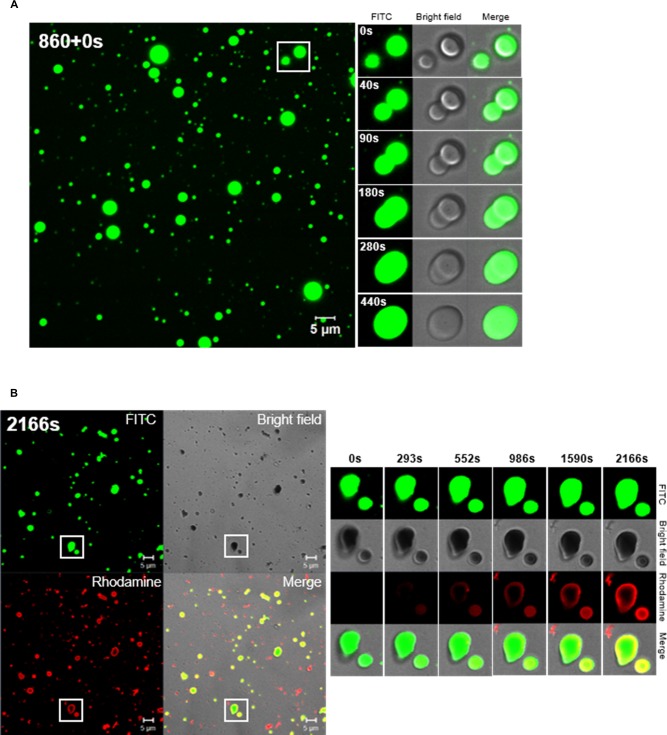
Fig 8. Formation of TP4 particles in the presence of sarkosyl.
(A) Fusion of TP4 particles at 0.5x Sar in a time-dependent manner. FITC-TP4 peptides (4 μg) were dissolved in 20 μl 0.5x Sar, loaded on cover glass and examined by CLSM 780. TP4 peptides existed in small particles and fused with each other. The images generated in the processing of TP4 particle fusion was taken at 860s after dissolving TP4 peptides in 0.5x Sar (within the white frame of left panel), and were also shown on the right with three fields (fluorescence, bright field and merged image). (B) Deposition of Rhodamine-TP4 peptides on existing FITC-TP4 particles. Rhodamine-TP4 peptides (2 μg in 20 μl 0.5x Sar) were added to a drop containing preformed FITC-TP4 particles (2 μg in 20 μl 0.5x Sar) on cover glass and examined by CLSM780. The Rhodamine-TP4 peptides existed in small/red particles by themselves or deposited on surrounding FITC-TP4 particles. The images at the right panel showed Rhodamine-TP4 deposition on existing FITC-TP4 particles taken at 2166s after the addition of Rhodamine-TP4 peptides at 0.5x Sar within the white frame of the left panel. The FITC-TP4, Rhodamine-TP4 and FITC-TP4/Rhodamine-TP4 complex are shown in green, red and yellow.

