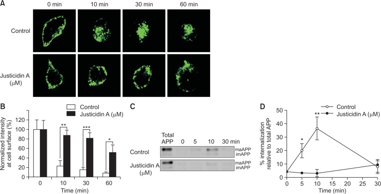
Fig. 4.
Endocytosis rate of APP was decreased by justicidin A. (A) Justicidin A decreased the endocytosis rate of APP. Cell surface APP was immunolabeled with 6E10 antibody in the presence of 1 µM justicidin A for 45 min at 4°C. Then, cells were incubated at 37°C for varying time periods, followed by fixation, and permeabilization. Cells were incubated with GFP-tagged secondary antibody and observed under a fluorescence microscope. (B) Fluorescence intensities of APP at the plasma membrane were obtained using Image J software from control (open bars) and justicidin-treated (closed bars) cells (n=10). (C) Cells were incubated with EZ-Link Sulfo-NHS-SS-Biotin at 4°C for 10 min. Next, cells were incubated with 1 µM justicidin A at 4°C for 45 min, followed by incubation at 37°C for various time periods. The remaining biotin at the cell surface was removed by reducing agent, and the internalized biotinylated proteins were pulled down using streptavidin beads. Representative Western blot shows the internalization of APP. (D) Internalized APP levels were quantified by the densitometric analysis of the bands. % of internalization was obtained by comparing to the total cell surface APP (n=5). *p<0.05, **p<0.01, ***p<0.001.

