Figure 1:

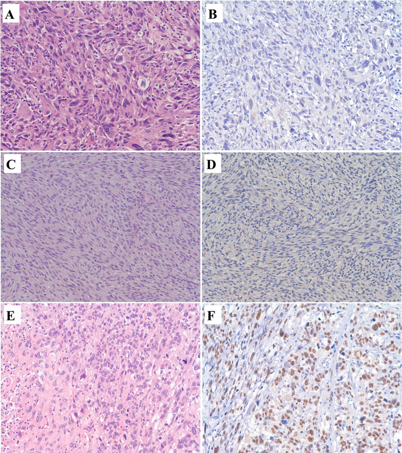
SMARCA1 immunohistochemistry in soft tissue sarcomas, A to F, Light Microscopy, 200x. A and B: Undifferentiated Sarcoma. A, Hematoxylin and Eosin: Undifferentiated sarcoma with haphazardly arranged and vaguely storiform, pleomorphic, anaplastic tumor cells with giant cells and hyperchromatic irregular nuclei. B. SMARCA1 Immunohistochemistry: Loss of expression of SMARCA1 in undifferentiated sarcoma. C and D: Malignant peripheral nerve sheath tumor. C, Hematoxylin and Eosin: Malignant peripheral nerve sheath tumor with tumor cells arranged in sweeping fascicles that are hypercellular, elongated nuclei and mitoses. D. SMARCA1 Immunohistochemistry: Loss of expression of SMARCA1 in malignant peripheral nerve sheath tumor. E and F: Leiomyosarcoma. E, Hematoxylin and Eosin: Leiomyosarcoma with tumor cells arranged in vague fascicles with eosinophilic cytoplasm, necrosis, enlarged hyperchromatic pleomorphic nuclei, and mitoses. F. SMARCA1 Immunohistochemistry: Nuclear expression of SMARCA1 in leiomyosarcoma.
