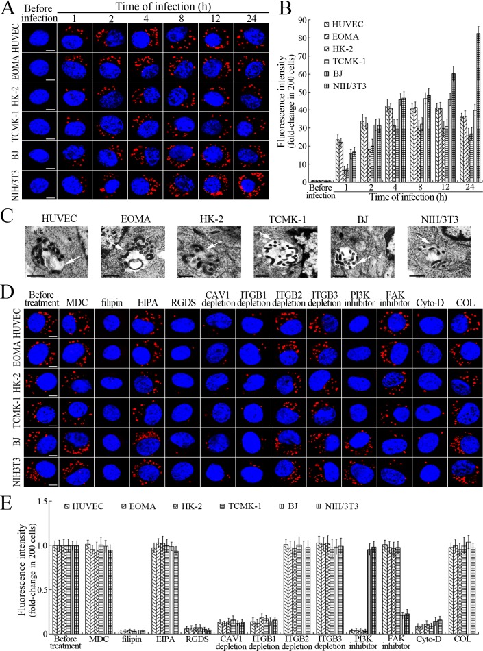
Figure 1. Internalization of L. interrogans into cells by CAV1/ITGB1-PI3K/FAK-mediated MF-dependent endocytosis.
(A) Leptospires in the cells infected with L. interrogans strain Lai for the indicated times, examined by confocal microscopy (scale bars = 5 μm). The blue plaques indicate the nucleus. The red spots around the nucleus indicate the intracellular leptospires. (B) Statistical summary of red fluorescence intensity reflecting the leptospires in the infected cells for the indicated times. Statistical data from experiments such as shown in (A). Bars show the means ± SD of three independent experiments. The red fluorescence intensity values from the cells without infection (before infection) were set as 1.0. (C) Lep-vesicles in the cells infected with L. interrogans strain Lai for 4 hr, examined by transmission electron microscopy (scale bars = 0.5 μm). The arrows indicate the intracellular leptospires in the membrane-bound vesicles. (D) Decrease of leptospires in the different inhibitor- or siRNA-treated cells infected with L. interrogans strain Lai for 4 hr, examined by confocal microscopy (scale bars = 5 μm). The legends are the same as shown in (A). (E) Statistical summary of red fluorescence intensity reflecting the leptospires in the different inhibitor- or siRNA-treated cells infected with L. interrogans strain Lai for 4 hr. Statistical data from experiments such as shown in (D). The other legends are the same as shown in (B).


