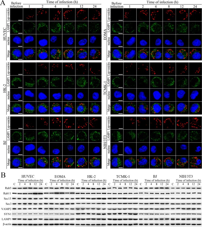
Figure 2. No fusion of Lep-vesicles with lysosomes and increase of target protein expression during infection.
(A) No co-localization of Lep-vesicles with lysosomes in the cells during infection with L. interrogans strain Lai, examined by confocal microscopy (scale bars = 5 μm). The blue plaques indicate the nucleus. The red or green spots indicate the Lep-vesicles or lysosomal marker LAMP1 molecules. No Lep-vesicle-LAMP1 co-localization (yellow) was found. (B) No significant expression increase of endocytic/excytotic and lysosomal proteins in the cells during infection with L. interrogans strain Lai, detected by Western Blot assay. C: the uninfected control cells.

