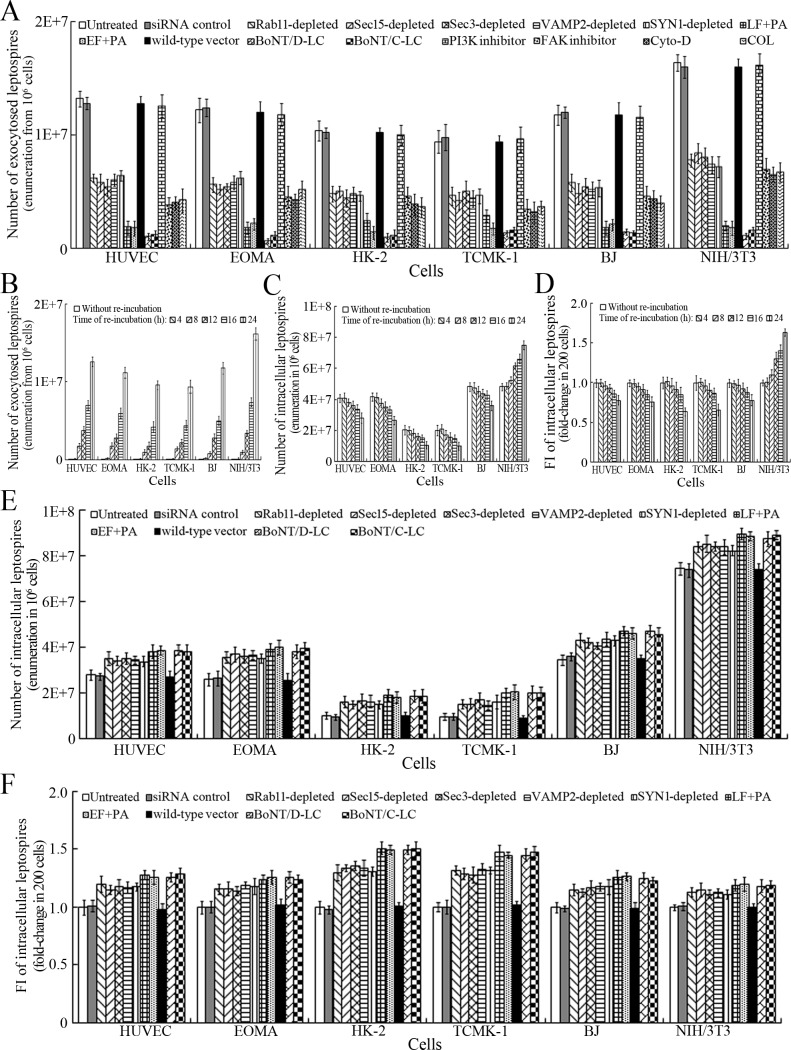
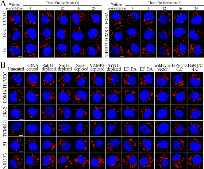
Figure 6. Exocytosis and propagation of intracellular L. interrogans.
(A) Endocytic recycling and vesicular transport systems-mediated exocytosis of L. interrogans strain Lai from the infected cells after a 24 hr extracellular leptospire-free re-incubation, examined by dark field microscopic Petroff-Hausser enumeration. Bars show the means ± SD of three independent experiments. (B) Release of L. interrogans strain Lai from the infected cells for the indicated times during extracellular leptospire-free re-incubation, examined by dark field microscopic Petroff-Hausser enumeration. Bars show the means ± SD of three independent experiments. (C) Number of L. interrogans strain Lai in the infected cells for the indicated times during extracellular leptospire-free re-incubation, examined by dark field microscopic Petroff-Hausser enumeration. The legend is the same as shown in (B). (D) Statistical summary of fluorescence intensity reflecting the number of L. interrogans strain Lai in the infected cells for the indicated times during extracellular leptospire-free re-incubation, examined by confocal microscopy. Bars show the means ± SD of three independent experiments. The fluorescence intensity values reflecting the leptospires in the cells after a 4 hr infection with the spirochete (without re-incubation) were set as 1.0. (E) Number of L. interrogans strain Lai in the siRNA- or toxin-treated infected cells after a 24 hr extracellular leptospire-free re-incubation, examined by dark field microscopic Petroff-Hausser enumeration. The legend is the same as shown in (B). (F) Statistical summary of fluorescence intensity reflecting the number of L. interrogans strain Lai in the siRNA- or toxin-treated infected cells after a 24 hr extracellular leptospire-free re-incubation, examined by confocal microscopy. The legends are the same as shown in (D).


