. 2019 Apr 23;8:e44594. doi: 10.7554/eLife.44594

Figure 9. Transcytosis of L. interrogans through cell monolayers mediated by endocytic recycling and vesicular transport systems.

(A) Decreased transcytosis of L. interrogans strain Lai through the filipin- or RGDS-inhibited cell monolayers, examined by transwell test and dark field microscopic Petroff-Hausser enumeration. The time points show the means ± SD of three independent experiments. (B) Decreased transcytosis of L. interrogans strain Lai through the Rab11-, Sec15-, Sec-3, VAMP2- or SYN1-depleted cell monolayers, examined by transwell test and dark field microscopic Petroff-Hausser enumeration. The legends are the same as shown in (A). (C) Decreased transcytosis of L. interrogans strain Lai through the LF + PA or BoNT/D-LC-treated cell monolayers, examined by transwell test and dark field microscopic Petroff-Hausser enumeration. The legends are the same as shown in (A).

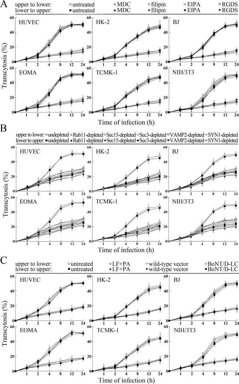
Figure 9.

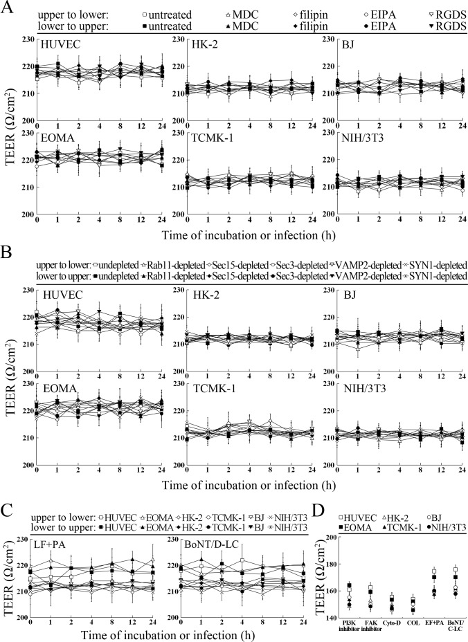
Figure 9—figure supplement 1. TEER values of cell monolayers in transwell test.

Figure 9—figure supplement 1.

(A) Higher TEER of the MDC-, filipin-, EIPA- or RGDS-treated cell monolayers during incubation or infection with L. interrogans for the indicated times, detected using a cell electric resistance indicator. (B) Higher TEER of the Rab11-, Sec15-, Sec3-, VAMP2- or SYN1-depleted cell monolayers during incubation or infection with L. interrogans for the indicated times, detected using a cell electric resistance indicator. (C) Higher TEER of the LF + PA or BoNT/D-LC-treated cell monolayers during incubation or infection with L. interrogans for the indicated times, detected using a cell electric resistance indicator. (D) Lower TEER of the LY294002-, 14/Y15-, Cyto-D, COL-, EF + PA or BoNT/C-LC-pretreated cell monolayers, detected using a cell electric resistance indicator.