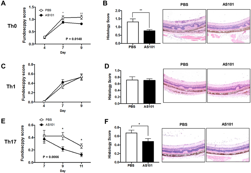
Fig. 5. AS101 suppresses EAU induced by IRBP-specific Th0 or Th17, but not Th1, effector T cells.
Cells were isolated from LN of CD90.1 R161H mice and stimulated with IRBP161-180 peptide under Th0, Th1 or Th17 conditions. After 3 days, viable cells were collected in Lympholyte M and adoptively transferred into CD90.2 B10.RIII WT mice. AS101 (27 ug, i.p.) was injected to the animals daily. (A) Disease was monitored by fundoscopy at days 4, 7 and 9 post-transfer of Th0 cells. Data pooled from 3 experiments. (B) Histology scores and representative images of H&E stained histology slides on day 10. (C) Fundoscopy scores of Th1 cell transferred mice. Data pooled from 6 experiments. (D) Histology scores and representative images of histology slides (H&E) on day 10. (E) Fundoscopy scores of Th17 cell transferred mice. Data pooled from 3 experiments. (F) Histology scores and representative images of histology slides (H&E) on day 11. Magnification; X100. Data shown as mean ± SEM. Significance was determined using Mann-Whitney test. *p<0.05, **p<0.01 vs. PBS group.

