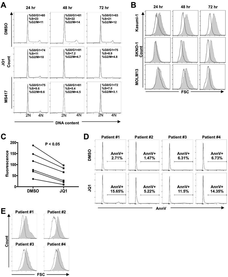
Figure 2. BETi induce cell cycle arrest and reduce cell size in t(8;21) AML cells.
(A) Cell cycle analyses of Kasumi-1 cells treated with BETi show cell cycle arrest with modest cell death 24–72 hr after treatment. Representative graphs of DNA content (2N to 4N) are shown (n=4). (B) Flow cytometry analyses showing forward scatter plots indicate that t(8;21) cells are distinctly smaller after treatment with 250 nM JQ1. Representative graphs are shown (n=4). Shaded area represents DMSO and white plots represent JQ1. (C and D) High blast count t(8;21) AML patient samples (n=6) were treated with 250 nM JQ1 for 3 days. AlamarBlue assays show the inhibition of cell growth p < 0.05 by two-sided Wilcoxon signed-rank test (C). (D) Shows the lack of Annexin V positive cells for four of the samples in C. (E) Forward scatter plots of flow cytometry analyses show that t(8;21) AML patient cells are distinctly smaller after JQ1 treatment for 3 days. Representative flow cytometry plots are shown. Shaded area represents DMSO and empty area represents JQ1.

