
Fig. 1.
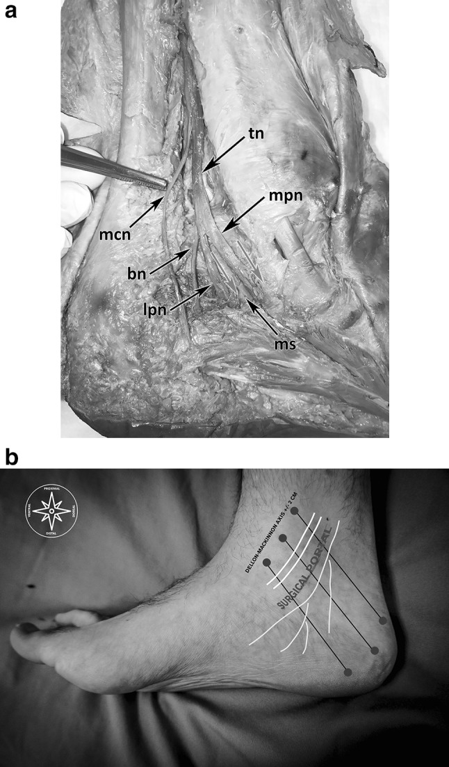
a Anatomical dissection of the tibial nerve and its branches running in the TT. tn Tibial nerve, mpn medial plantar nerve, lpn lateral plantar nerve, bn Baxter nerve, mcb medial calcaneal branch, ms medial intermuscular septum, red arrows: nerves entering separated tubes. b The red circles show the landmarks to sketch the flexor retinaculum; the black lines show the DM-line and the lines ± 2 cm (proximal and distal) to the DM-line; the yellow sketched lines show course of the tibial nerve with its branches; the white lines show the courses of the posterior tibial and the flexor digitorum muscles. (Color figure online)