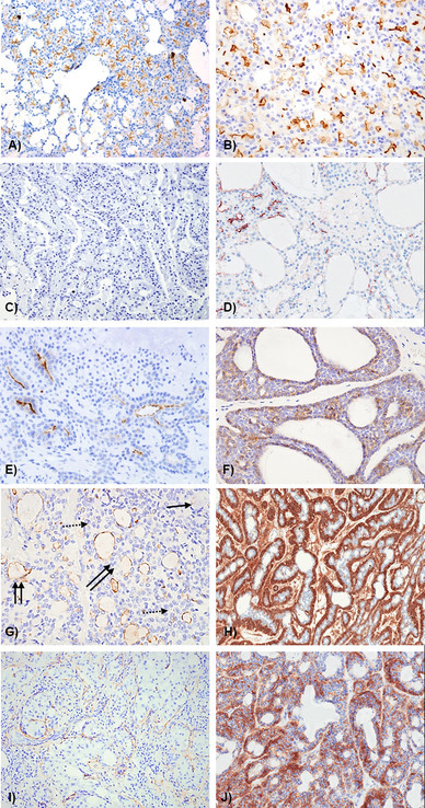
Fig. 1.

Representative photomicrographs showing DOG-1 staining pattern in a normal gland; b acinic cell CA; c secretory carcinoma—negative; d secretory carcinoma—focal positive, e pleomorphic adenoma; f adenoid cystic CA; g mucoepidermoid carcinoma; h basal cell adenoma; i carcinoma in pleomorphic adenoma; j papillary cystadenocarcinoma. g Solid arrow—mucous cells, dotted arrow—intermediate cells, double arrow—luminal spaces with DOG1 positivity
